MRI After LEEP in Low-Risk Cervical Cancer: SHAPE Findings
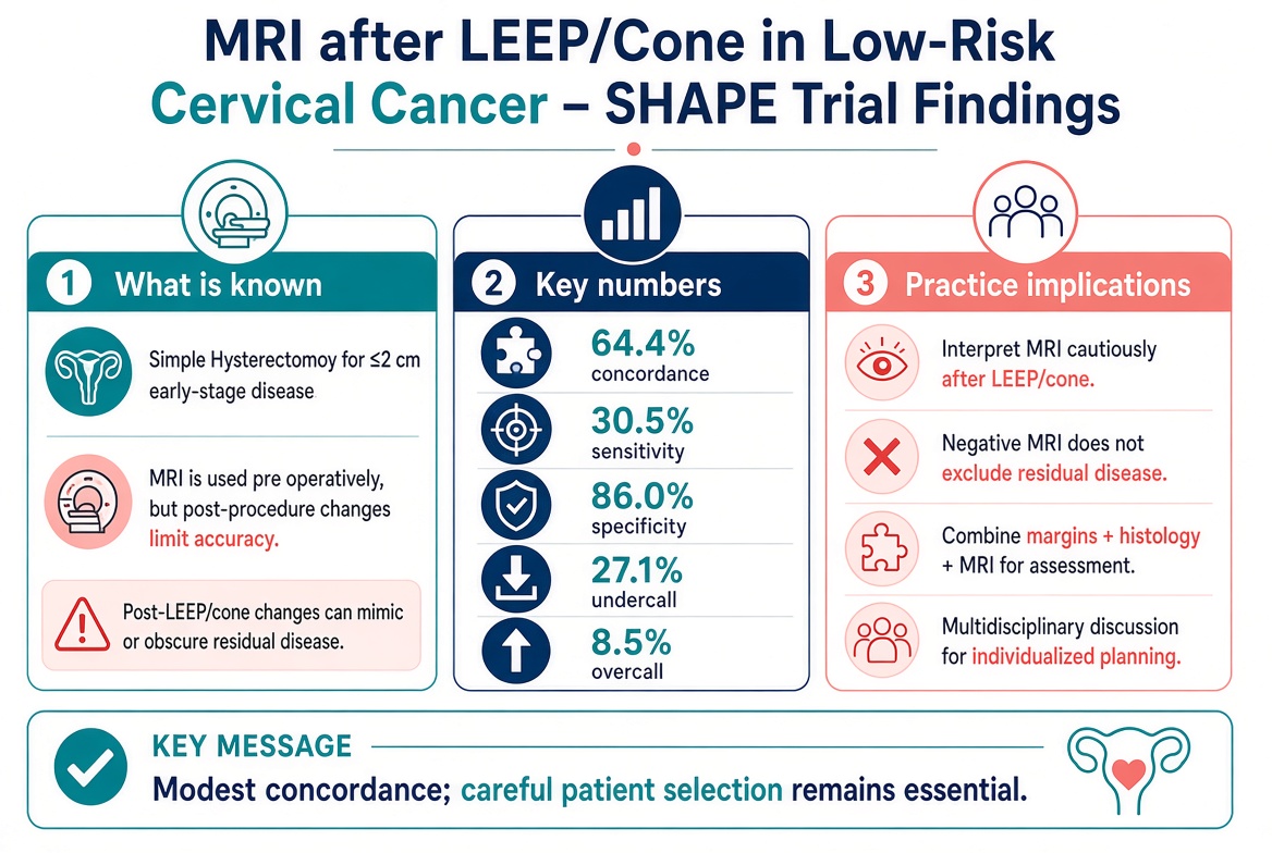
An exploratory analysis of the SHAPE trial found 64.4% concordance between pelvic MRI after LEEP/cone and final hysterectomy pathology in low-risk cervical cancer, with only 30.5% sensitivity for residual disease. Negative MRI does not exclude residual tumor; margin status and multidisciplinary review remain essential.

GLOBAL NEWS · RESEARCH BRIEFING
MRI After LEEP or Cone Biopsy in Low-Risk Cervical Cancer: What the SHAPE Trial Exploratory Analysis Shows
Modest concordance between post-procedure MRI and final hysterectomy pathology, with practical implications for surgical planning
For publication on gynecology.hospital Editorial Team · 14 October 2026 · Category: Global News / Cervical Cancer |
The SHAPE trial (CX.5) established that simple hysterectomy is non-inferior to radical hysterectomy for carefully selected women with low-risk early cervical cancer (FIGO 2009 stage IA2–IB1, tumors ≤2 cm with limited stromal invasion). An exploratory analysis published in the International Journal of Gynecological Cancer examined how well pelvic MRI performed after diagnostic LEEP or cone biopsy correlated with final hysterectomy pathology in that same cohort. The findings are relevant for any center that relies on post-excision imaging to confirm eligibility for less radical surgery.
What the SHAPE trial established
SHAPE randomized approximately 700 patients with low-risk disease to simple or radical hysterectomy plus lymph-node assessment. With a median follow-up of 4.5 years, the 3-year pelvic recurrence rate was 2.52 percent after simple hysterectomy and 2.17 percent after radical hysterectomy—an absolute difference well within the non-inferiority margin. Simple hysterectomy also produced fewer urinary complications. Eligibility required either limited invasion on the diagnostic LEEP/cone specimen or less than 50 percent stromal invasion on preoperative MRI, plus no evidence of nodal metastasis. Many patients therefore arrived at surgery having already undergone an excisional procedure whose margins and residual disease status influenced both imaging interpretation and final treatment decisions.
MRI after LEEP or cone: the exploratory analysis
Among 480 patients who had pelvic MRI after diagnostic LEEP or cone, concordance between MRI and final hysterectomy pathology was 64.4 percent (95 percent CI 59.5–68.7). Sensitivity for residual disease was only 30.5 percent and specificity 86.0 percent. The undercall rate—MRI showing no disease when residual disease was present—was 27.1 percent; the overcall rate was 8.5 percent. Concordance was higher when LEEP/cone margins were negative (76.6 percent) than when they were positive (57.3 percent). Adenocarcinoma had a lower positive predictive value (41.9 percent) than squamous histology (66.2 percent).
Key quantitative findings:
64.4 percent overall MRI–pathology concordance.
30.5 percent sensitivity and 86.0 percent specificity for residual disease.
27.1 percent undercall rate; 8.5 percent overcall rate.
62.5 percent of lesions that measured ≥2 cm on final pathology were not seen on MRI.
Ten patients had no disease on MRI yet tumors >2 cm on final pathology; six of them underwent simple hysterectomy.
Forty-one patients had disease on MRI but no residual disease on final pathology; twenty underwent radical hysterectomy.
Clinical implications
The analysis concludes that concordance is only modest and that the impact on under- and over-treatment rates was limited within the trial population. Nevertheless, the low sensitivity means a negative MRI after LEEP or cone does not reliably exclude residual disease, particularly when margins are positive. Post-procedure inflammation and tissue distortion can both obscure and mimic residual tumor. In practice this supports a cautious reading of post-LEEP MRI and reinforces the value of integrating margin status, histology, and imaging in a multidisciplinary discussion before committing to simple versus radical hysterectomy.
Interpret post-LEEP/cone MRI with caution; a negative study does not exclude residual disease.
Combine LEEP/cone margin status, histologic subtype, and MRI findings rather than relying on any single modality.
Inform radiologists of the exact procedure performed and the interval since excision, as these details affect interpretation.
Reserve simple hysterectomy for patients who clearly meet low-risk criteria on both pathology and imaging; when doubt remains, consider more radical surgery or further assessment.
Future work on radiomics or refined imaging protocols may improve selection, but current evidence does not support routine de-escalation based on a negative post-LEEP MRI alone.
For clinicians managing early cervical cancer, the SHAPE trial supports less radical surgery in well-selected patients, but the exploratory MRI analysis underscores that imaging after diagnostic excision is an imperfect tool. Margin status and careful clinicopathologic correlation remain central to safe decision-making.
Sources
Plante M et al. Correlation of pelvic magnetic resonance imaging with diagnostic loop electrosurgical excision procedure/cone biopsy and hysterectomy pathology: an exploratory analysis of the CX.5/SHAPE trial. Int J Gynecol Cancer. 2026.
Plante M et al. Simple versus Radical Hysterectomy in Women with Low-Risk Cervical Cancer. N Engl J Med. 2024;390:819-829.
Canadian Cancer Trials Group CX.5/SHAPE trial publications and exploratory analyses on surgical approach and LEEP/cone margins.
Research briefings by the gynecology.hospital editorial team summarise published studies and official data, and list their sources at the end of each article.